Skip to main
![]() Scroll to content
Scroll to content

104th Annual Meeting
April 27 - 30, 2024 at the Metro Toronto Convention Center, Toronto, ON, Canada
The world's most prestigious thoracic surgery event. Grow your expertise, network with leading experts, and help build the future of thoracic surgery.
In Collaboration With:
Join Us in Toronto
Join Us in Toronto
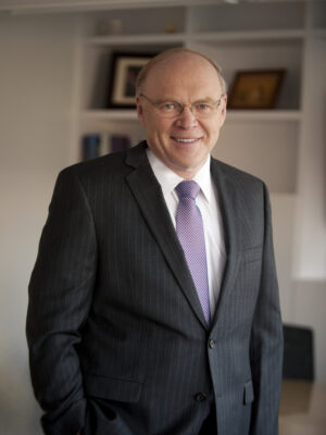
On behalf of my excellent Program Committee, I am thrilled to invite you to join us for the AATS 104th Annual Meeting in Toronto, April 27-30. Toronto has been a wonderful host for our meetings in years past. We are coming off an incredible 103rd Annual Meeting led by our first woman president, Dr. Yolonda L. Colson, and I am looking forward to building upon that energy when we head to Toronto in the Spring 2024.
We had a record-setting number of abstracts and case video submissions this year, which will undoubtedly lead to a dynamic, cutting-edge program with the best cardiothoracic surgery educational offerings in our specialty. We added a new submission category focused on structural heart which will add to the already extraordinary variety of educational features we have in place for the program. In the afternoon, we will have non-CME cutting-edge presentations on new innovations, inventions, and pioneering research in our specialty.
It is an honor to be selected to serve as the AATS President. I do not take this leadership role lightly, and therefore envision a meeting where we can all learn valuable lessons of leadership no matter what stage we are at in our career paths. Leadership is a skill and value we build upon and develop throughout our professional journeys. Throughout the planning stages of this meeting, we’ve made significant efforts to highlight programmatic elements focused on discussions and collaborations to advance our leadership skills, including a theorem for leadership you can build on. We also trust you will find this meeting will contribute to your academic endeavors and surgical skills.
Read More
Marc Gillinov
Since joining the Cleveland Clinic in 1997, A. Marc Gillinov has become one of the nations busiest heart surgeons. He is the Chair of the Department of Thoracic and Cardiovascular Surgery and specializes in robotic and minimally-invasive heart valve repair and replacement. His patients range from Academy Award winner Robin Williams to his long-time barber, Vince.
See the Full Planning Committee

Y. Joseph Woo
Joseph Woo, M.D. serves as the Norman E. Shumway Professor and Chair of the Department of Cardiothoracic Surgery at Stanford University and holds a courtesy appointment in the Department of Bioengineering. He received his undergraduate degree from the Massachusetts Institute of Technology and his M.D. from the University of Pennsylvania where he also conducted his postgraduate surgical training in general surgery and cardiothoracic surgery as well as a postdoctoral research fellowship developing novel molecular strategies for attenuating myocardial ischemic injury.
See the Full Planning Committee

Industry Programming
In addition to the AATS Annual Meeting accredited program AATS industry partners will host a dynamic range of sessions, available to all registered attendees.
View Industry SessionsComplex Valve Scenarios Symposium
Learn More
The AATS Complex Valve Scenario Symposium will gather leading surgeons and interventionalists from around the world to share their approaches to the most complicated problems in the field of heart valve disease. This half-day symposium presents a focused, unique opportunity to learn from the masters of heart valve disease.